See Explanation
[ "https://i.postimg.cc/HkFwB1Wn/case-212816-image-1-1024px-diabetic-charcot-foot-deformity.jpg" ]
Orthopedics
miscellaneous
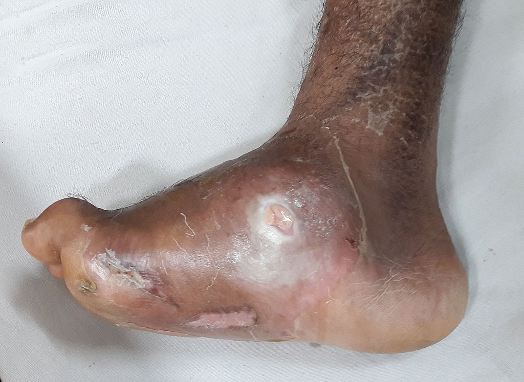
neuropathic (charcot) arthropathy
A 60-year-old woman with a long-standing history of type 2 diabetes mellitus, complicated by diabetic retinopathy and peripheral neuropathy, presents to an outpatient clinic. She reports a persistent, non-tender ulcer on the medial plantar aspect of her left foot, which she first noticed approximately 8 months ago. Despite attempts at wound care and occasional use of special shoes, the ulcer intermittently reopens, especially after prolonged walking. She denies any significant pain associated with the ulcer or the foot deformity itself. Her medical history also includes hypertension, hyperlipidemia, and a prior episode of atrial fibrillation. Her current medications include metformin, insulin glargine, empagliflozin, valsartan, gabapentin, and rosuvastatin.On physical examination, her left foot appears warm to touch with moderate, non-pitting edema extending to the ankle. There is a noticeable collapse of the longitudinal arch, giving the foot a "rocker-bottom" appearance. A 2 cm x 3 cm ulcer with a clean base is noted on the medial plantar surface. Monofilament testing reveals absent sensation in the distal left foot, and vibratory sensation is significantly diminished at the left ankle. Pulses (dorsalis pedis and posterior tibial) are palpable but diminished. Capillary refill time is less than 3 seconds. Musculoskeletal examination reveals preserved range of motion at the ankle and toes, but midfoot instability.Which of the following represents both the most likely underlying condition contributing to this foot deformity and the most appropriate immediate management strategy?
| Lab Parameter | Value | Reference Range |
|---|---|---|
| Hemoglobin A1c | 9.8 % | < 6.5 % |
| White Blood Cell Count | 7.2 x 10^9/L | 4.0-11.0 x 10^9/L |
| C-reactive protein | 5 mg/L | < 3 mg/L |
Edit question